Bhopal Aerosol Tragedy - Physical Findings Naked Eye Illustrated Graphically
 |
 |
 |
 |
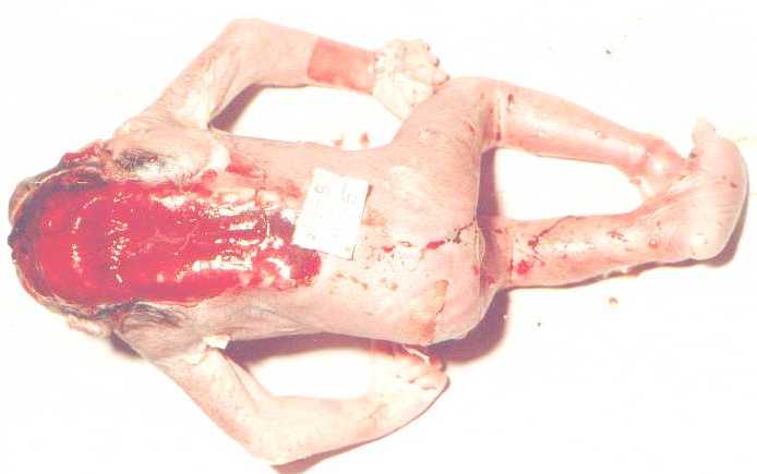 |

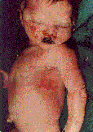
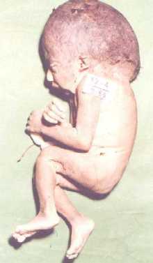
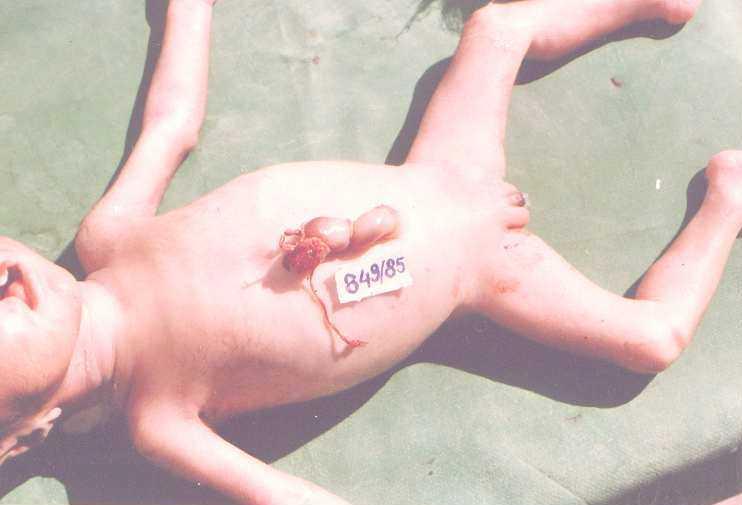
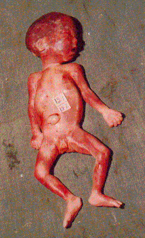
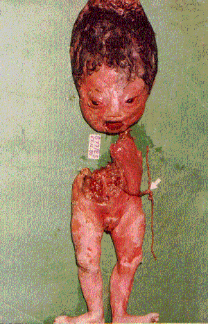
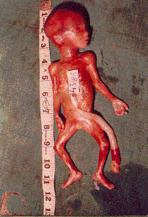
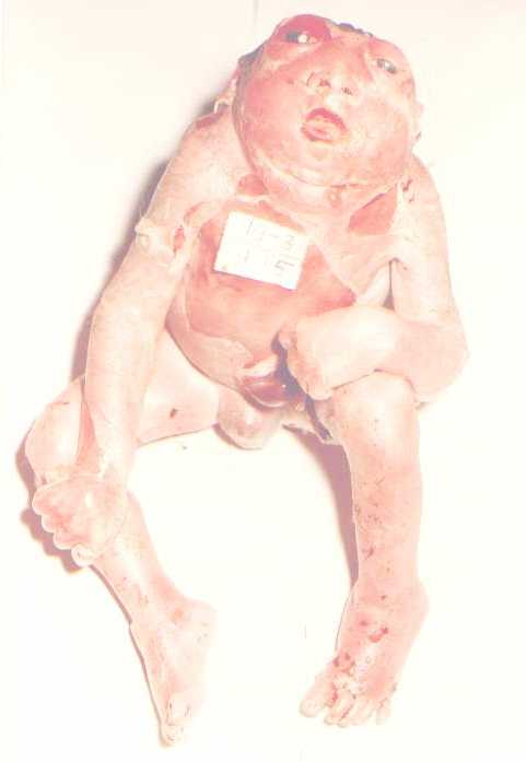
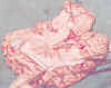
(4) - Ventral and dorsal view of enancephaly bany norn after about 10 months of the eposure o the mother, (5) - The dorsal view opened, (8) - Baby hydranemous born 10 month after the tragedy, this period showed many congenital anomaly, (9) - A child recently born showing pink coloration of the body with umbilical herniation and hair lip with cleft pallet
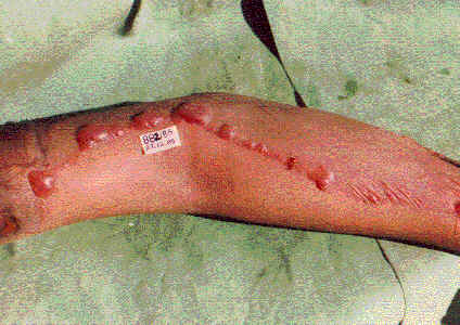
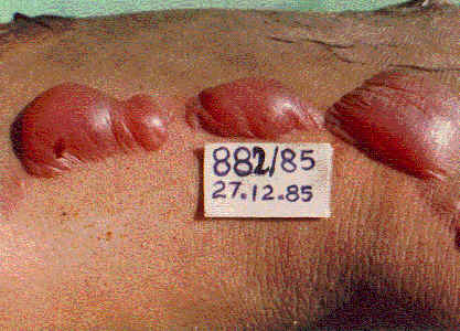
Reaction Of Gases on Skin
 |
 |
 |
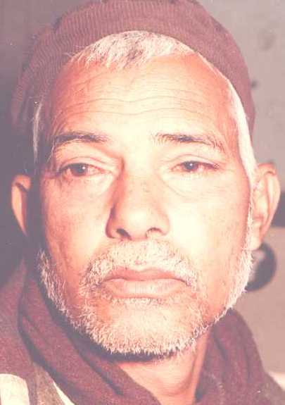
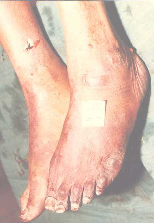
(3) - The exposed part of neck and chest shows the reaction of the skin from Aerosol, (4) - Exposed Victims showing Pink Coloration in the face with congested eyes, (5) - The legs show the reaction of the skin to the exposure of the Aerosol with red coloration all round. Hypostasis not limited to dependent parts,
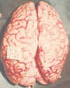
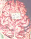
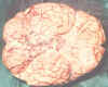
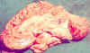
Brain
 |
 |
 |
 |
 |
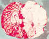 |
Brain surfaces showing stasis in the blood vessels, which is characterised by the absence of venous coloration. The medial surface characterised by the absence of cerebrum shows the same feature with generalised oedema. The vessels have become very prominent with increased viscosity resulting in congestion and pattern appearances. Softening of the white matter was a common feature. These photographs nearly relate 12 weeks period from the day of disaster.
Lever
 |
 |
 |
 |
 |
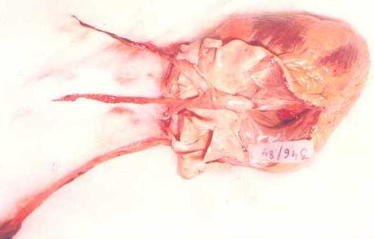
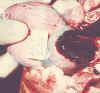
(1,2) - External surface of the lever showing petechial and eneralaxed hemorrhages with distended gall bladder - a common finding, and having a extricating in the cut surface, (3,4,5) -The external surfaces of the liver showing oetechial and generalized hemorrhages with distended gall bladder - a common finding, and having a extict tinge in the cut surface, (6) - After about two months, changes in the blood showing various coloration appeared but from the arterial stresses with petechial and generalized hemorrhages seen.
Lungs
(1) - Intense hemorrhages congestion of the trachea with absence of venous coloration, (2) - Nearly two months after; dark clotted blood seen on the vessels of the hylum of the lings with surfaces covered with petechial and generalised hemorrhages, (3) - Another Autopsy showing lungs with the same features as(2), (4)- Hemorrhages of the surface of the lungs more in the basal region but all around, cherry red in color, the interlobular region are more hemorrhage


Kidney

Kidneys showing generalized hemorrhages on the surface with localized petechial hemorrhages
Autopsy Findings
 |
 |
(1)-Autopsy Findings in 1984 cases (Total Cases394) (2) - Autopsy Findings in 1985 (Total Cases 105)
Heart
 |
 |
 |
 |
 |
 |
(1) - Hemorrhages seen on the surface of the heart with coagulated blood in the great blood vessel as a cast, (2) - Another Heart 6 days after the exposure showing hemorrhage myocardium with petechial hemorrhage and also focal hemorrhage, (3) - The First autopsy of 21st December 1984 showing apical petechial hemorrhages, (4) - External surface of the heart showing the generalized hemorrhage with scattered petechial hemorrhages, (5) - Cast of coagulum in the great blood vessels of the heart.
Stomach
 |
 |
 |
 |
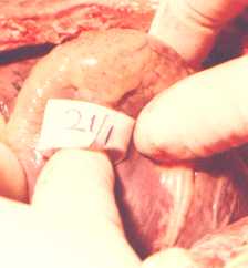
(1) - Photograph shows thoracic and abdominal cavity with vessels heavily congested with arcades and lover, spleen, showing haemirrhagic surfaces. (2) - A photograph showing the entire thoracic and abdominal cavity with chocolate like liver due to hemorrhage, generalized hemorrhage in the lings and congested in the vessels the intestine, (3) - The intense congestion of the stomach mucosa even after one month of the tragedy, (4) - Hour glass hemorrhage appearance of the stomach contents clearly demarcated